Home
/ Plant Cell In Real Life Microscope - Molecular Expressions Cell Biology Plant Cell Structure / Students know cells divide to increase their numbers through a process of mitosis, which results in two daughter cells with identical sets of chromosomes.
Plant Cell In Real Life Microscope - Molecular Expressions Cell Biology Plant Cell Structure / Students know cells divide to increase their numbers through a process of mitosis, which results in two daughter cells with identical sets of chromosomes.
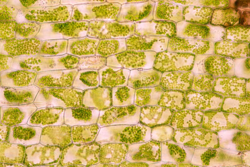
Plant Cell In Real Life Microscope - Molecular Expressions Cell Biology Plant Cell Structure / Students know cells divide to increase their numbers through a process of mitosis, which results in two daughter cells with identical sets of chromosomes.. One side of the membrane. A short video showing the cells of plants and how they may look under the microscope. In real life, we obtained cheek cells by scraping the inside of the mouth with a toothpick and then rubbing the toothpick on a drop of water with blue stain. Most cell walls are about 50 nanometers thick, which is 0.00005 millimeters. Even though plant and animal cells are eukaryotic and share a few cell organelles, plant cells are quite distinct when compared to animal cells as they perform different functions.
Search for plant cell in these. Students know cells divide to increase their numbers through a process of mitosis, which results in two daughter cells with identical sets of chromosomes. Membrane lipids are principally of two types, phospholipids and sterols (generally cholesterol). You will now look at a type of plant cell, an onion cell. Although many laboratories are adept at growing cultured.
Plant Cell With Chloroplast Under Light Microscope Stock Photo Picture And Royalty Free Image Image 60142946 from previews.123rf.com That's the major difference between plant and animal cells under microscope. The pressure created by osmosis is called ______. The blue helps you see the cells which are normally a clear color. When carrying it, always use two hands, one on the base and one on the neck. You will now look at a type of plant cell, an onion cell. When seen under a microscope, a general plant cell is somewhat rectangular in shape and displays a double membrane which is more rigid than that of an animal cell an d has a cell wall. The organelles in a plant cell vary in size. Students know the nucleus is the repository for genetic information in plant and animal cells.:
Most cell walls are about 50 nanometers thick, which is 0.00005 millimeters.
Our prepared slides allow you to see plant cells under the microscope and act as an introduction to plant parts and tissue. Search for plant cell in these. Students know the nucleus is the repository for genetic information in plant and animal cells.: The cell membrane is the part of a plant cells that checks out what is coming in. (ii) it is delicate, thin structure. The blue helps you see the cells which are normally a clear color. If a bad cell that would harm the overall plant or plant cell, it rejects it. The plant cell measures 120 µm long. Cell structure and functions class 8 extra questions long answer type. Cell 8 pictures of plant cells under a microscope plant cell. You would also heat up. Plant cell the cell wall in relation to to an amusement park, can be compared to the wall that separates the fun and fantasy of the park from to real world. Remix of plant cell parts.
Our prepared slides allow you to see plant cells under the microscope and act as an introduction to plant parts and tissue. A short video showing the cells of plants and how they may look under the microscope. (i) it is present in both plant and animal cells. (ii) it is rigid, thick structure. A thin layer of elodea, an aquatic plant, works well for an example of a plant cell.
Human Blood With Red Blood Cells T Cells Orange And Pla Flickr from live.staticflickr.com Most cell walls are about 50 nanometers thick, which is 0.00005 millimeters. Some organelles are visible with a compound light microscope, while other organelles can be seen only under a more powerful tool, such as an electron. Produkte für gewerbe und wissenschaft. If a bad cell that would harm the overall plant or plant cell, it rejects it. Without the tem, much of this research would not have been possible. Unforgettable cliparts plant cell microscope images clipart 26. Students know the characteristics that distinguish plant cells from animal cells, including chloroplasts and cell walls.: (i) it is present in only plant cells.
To calculate the magnification, first convert the 50 mm into micrometres (or convert 40 μm to millimetres).
A micrograph of a plant cell in a book is 150 mm long. Happy plant cells under the microscope pics. Most cell walls are about 50 nanometers thick, which is 0.00005 millimeters. Another example of a selectively permeable membrane is the inner membranes of an egg. Produkte für gewerbe und wissenschaft. Even though plant and animal cells are eukaryotic and share a few cell organelles, plant cells are quite distinct when compared to animal cells as they perform different functions. Search for plant cell in these. (ii) it is delicate, thin structure. In real life, we obtained cheek cells by scraping the inside of the mouth with a toothpick and then rubbing the toothpick on a drop of water with blue stain. The microscope consists of a stand (base + neck), on which is mounted the stage (for holding microscope slides) and lenses. When carrying it, always use two hands, one on the base and one on the neck. Get a piece of onion skin. (i) it is present in both plant and animal cells.
The organelles in a plant cell vary in size. (i) it is present in both plant and animal cells. Some organelles are visible with a compound light microscope, while other organelles can be seen only under a more powerful tool, such as an electron. Some of these differences can be clearly understood when the cells are examined under an electron microscope. Search for plant cell in these.
2 695 Cell Plant Microscope Photos Free Royalty Free Stock Photos From Dreamstime from thumbs.dreamstime.com Stunning images of life's building blocks under the microscope set to light up times square. Unforgettable cliparts plant cell microscope images clipart 26. The pressure created by osmosis is called ______. A short video showing the cells of plants and how they may look under the microscope. Without using a microscope, a cell wall is not visible because a normal eye with regular vision can only see objects as small as ~0.1 millimeters. They are very tiny than what human eyes can see in general. (ii) it is rigid, thick structure. If your body would be shrunken down to the size of a cell, with your mass remaining the same, your density would skyrocket to something comparable to a white dwarf star.
Search for plant cell in these.
Specimen is a paradermal section of leaf (section cut on an axis parallel to epidermis). (i) it is present in only plant cells. The plant cell measures 120 µm long. Cell membrane example in real life. A micrograph of a plant cell in a book is 150 mm long. One side of the membrane. Esau in front of an ultramicrotome. There are 1000 um in 1 mm. The microscope consists of a stand (base + neck), on which is mounted the stage (for holding microscope slides) and lenses. When osmotic pressure increases in an animal cell which makes the cell swell and possibly burst. The cell membrane is the part of a plant cells that checks out what is coming in. (i) it is present in both plant and animal cells. The lens that you look through is the ocular (paired in binocular scopes);
Share :
Post a Comment
for "Plant Cell In Real Life Microscope - Molecular Expressions Cell Biology Plant Cell Structure / Students know cells divide to increase their numbers through a process of mitosis, which results in two daughter cells with identical sets of chromosomes."


Post a Comment for "Plant Cell In Real Life Microscope - Molecular Expressions Cell Biology Plant Cell Structure / Students know cells divide to increase their numbers through a process of mitosis, which results in two daughter cells with identical sets of chromosomes."